Introduction
Teeth represents as an useful material for age estimation. The development of an individual can be affected by genetic, nutritional, hormonal and environmental factors.[1] But, it has been reported that dental mineralization is less affected by external factors when compared to skeletal mineralization.[1]
Teeth, being the hardest calcified tissue of the body tend to remain intact even when the other components of the skeletal system get disintegrated. The high resistance of the teeth to severe insults such as heat, cold and chemical agents makes them the most preferable tissue for forensic studies.[6]
The dental age can be assessed in young children with greater accuracy because many teeth will be undergoing development and calcification at the same time.[3],[4],[5] After the early teens most of the teeth get calcified and erupt except for the third molars. This makes the third molars the most important teeth of choice for age estimation in the late teens and early twenties.[2],[4]
Although the third molars might show some amount of variation in their anatomy and position, several authors state that are the most reliable biologic indicator for estimation of age during the middle teens and early twenties especially when there is a need to determine the juvenile or adult status of an individual when no valid document is available.[5],[6]
Numerous methods have been proposed to estimate the age using third molars. Modified Demirjian technique is an excellent method for age estimation from the radiological appearance of the third molar. The aim of this study was to categorize the orthopantamographs collected from the 515 children and young adults, according to the developmental stages proposed by modified Demirjian technique and to correlate it with the chronological age and sex of the subjects.
Material and Methods
This study includes 515 orthopantomo graphs of the subjects within the age group of 8 to 23 years (273 males and 242 females), from the Department of Orthodontics, Vyas Dental College and Hospital, in the period from 2000- 2011. Selection criteria included the following: Indian, well nourished, normal growth and development, and free of any known serious illness. The radiographs were examined separately by two observers. Examination and classification covered the development phase of right third molar and when not present, the contralateral molar was considered. Tooth calcification was rated according to the Modified Demirjian method in which one of the eight stages of calcification A to H, was assigned to the third molar tooth (Figure 1).
![Figure 1: Schematic drawing of the eight stages of crown - root formation of the molars according to modified Demirjian technique.[3]](article-image-3311-FIGURE_1_SCHEMATIC_DRAWING_OF_THE_EIGHT_STAGES_OF_.jpg) | Figure 1: Schematic drawing of the eight stages of crown - root formation of the molars according to modified Demirjian technique.[3]
![Figure 1: Schematic drawing of the eight stages of crown - root formation of the molars according to modified Demirjian technique.[3]](images/article-image-enlarge.jpg) |
Stage A: Calcification of single occlusal points without fusion of different calcifications.
Stage B: Fusion of mineralization points; the contour of the occlusal surface is recognizable.
Stage C: Enamel formation has been completed at the occlusal surface, and dentine formation has commenced. The pulp chamber is curved, and no pulp horns are visible.
Stage D: Crown formation has been completed to the level of the cemento-enamel junction. Root formation has commenced. The pulp horns are beginning to differentiate, but the walls of the pulp chamber remain curved.
Stage E: The root length remains shorter than the crown height. The walls of the pulp chamber are straight, and the pulp horns have become more differentiated than in the previous stage. Radicular bifurcation has commenced to calcify.
Stage F: The walls of the pulp chamber now form an isosceles triangle, and the root length is equal to or greater than the crown height. The bifurcation has developed sufficiently to give the roots a distinct form.
Stage G: The walls of the root canal are now parallel, but the apical end is partially open.
Stage H: The root apex is completely closed (distal root in molars). The periodontal membrane surrounding the root and apex is uniform in width throughout.
The first four stages (A–D) show crown formation from the beginning of cusp calcification to completed crown, and the second four (E– H) root formations from initial radicular bifurcation to apical closing. Descriptive statistics were obtained by calculating the means, standard deviations, and range of the chronologic ages for the eight stages of dental development. Statistical analysis was performed using the Mann-Whitney test between the age and sex.
Results
Totally 515 subjects were included in the study among which 273 (53%) were males and 242 (47%) were females. The interobserver agreement for the males: 95%, for females: 92% and the overall: 94%. The Mean, Standard Deviation, standard error for Mean, other descriptive statistics and Median for modified Demirjian stages of males (Table 1). The Mean, Standard Deviation, standard error for Mean, other descriptive statistics and Median for modified Demirjian stages of females (Table 2).
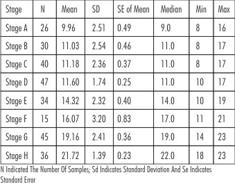 | Table 1: Descriptive Statistics For The Modified Demirjian Stages Of Males
|
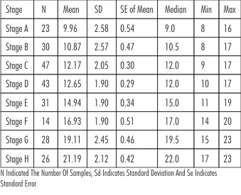 | Table 2: Descriptive Statistics For The Modified Demirjian Stages Of Males
|
The difference in mean age between males and females was found to be statistically significant regarding the calcification of Stage C (P<0.05) and Stage D (P<0.01). In addition no significant difference in mean age was found between males and females in Stage A, Stage B, Stage E, Stage F, Stage G and Stage H (P>0.05) (Table 3). The comparison between both the genders according to modified Demirjian stages: mean age and distribution of age is depicted in (Figure 2).
 | Table 3: Descriptive Values And Statistical Comparisons Of Modified Demirjian Stages In Both Sexes
|
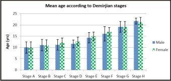 | Figure 2: Shows The Mean Age According To Modified Demirjian Stages In Both The Sexes
|
Discussion
Age estimation is an important exercise for medico-legal (age at death, criminal law cases, legal matters, adult status establishment, etc) and clinical purposes.[3],[6],[7] The need for age estimation has certain important reasons at certain specific age groups: a) 12 years: children below this age are not liable for certain offences; b) 14 years: a child cannot be employed below 14 years; c) 18 years: determines the status of majority and the legally permissible age of marriage in females; d) 21years: legally permissible age of marriage in males.[8] Numerous methods have been proposed for determination of the age. Studies have shown that dental development relates more closely to chronological age than skeletal, somatic or sexual maturity indicators. Tooth formation has been more widely used than tooth eruption for assessing dental maturation, because it is a continuous and progressive process. The methods of age estimation in which tooth is used as an evaluation tool include both – methods requiring the extraction of teeth and Histopathological examination and in-vivo methods which merely require radiographs.[8],[9]
One of the most widely used method is that of Demirjian, Goldstein and Tanner, which was first described in 1973 based on a large number of French– Canadian children.[10] The method evaluates the development of seven mandibular teeth from a panoramic radiograph and calculates dental age. Each tooth is rated according to developmental criteria (amount of dentine deposition, shape changes of pulp chambers, etc.) rather than the changes in size. Eight stages A to H, were defined from the 1st appearance of calcifications to the closure of apex and each one is allocated a score. The sum of those scores for an individual provided an estimate of dental maturity on a scale (overall maturity scores). These scores are subsequently converted into a dental age using conversion tables.[9]
Third molar development was not included in their investigations. It was only Moores et al and Schour & Masslef and the detailed work by Mincer et al that provided a favorable study of the age related stages of development of third molars.[2]
Numerous authors have conducted studies using modified Demirjian’s classification and indicated that reproducibility is higher for mandibular third molars than maxillary third molars.[9]
In the present study we assessed the developmental stages of third molar and compared it with the chronological age and sex of the subjects. 515 subjects were included in the study, the orthopantomographs of the subjects were collected and they were evaluated by modified Demirjian method and the appropriate stage was assigned.
In the present study the Mean age of 9.96years (Median: 9years) was observed for both the genders with respect to stage A, for the Stage B: Mean age of 11.03years for males and 10.87years for females (Median: males – 11years; females- 10.5years), for Stage C: Mean age of 11.18years for males and 12.17years for females(Median: males- 11years; females- 12years), For Stage D: Mean age of 11.60years for males and 12.65 years for females (Median: males- 11years; females- 12years), for Stage E: Mean age of 14.32years for males and 14.94years for females (Median: males- 14years; females- 15years), for Stage F: Mean age of 16.07 years for males and 16.93 years for females (Median: males-17years, females- 17years), for Stage G: Mean age of 19.16 years for males and 19.11years for females (Median: males- 19 years; females- 19years) and for Stage H: Mean age of 21.72 years for males and 21.19 years for females(Median: males- 22years; females- 22years).
Based on our results it can be assumed the mandibular third molars development starts at the similar age in both the sexes based with respect to the results of Stage A, but with respect to the further stages such as B, C, D, E and F the development is comparatively about 6 months to 1 year earlier in males than in females. But in the stages G and H the development of thirds molars are faster in females than in males by about 5-6 months.
As concluded by Mincer et al. in the A.B.F.O study, the examination of third molars may provide reasonable accuracy for the likelihood that a person is at least, e.g., 18 years old, instead of the estimation of exact chronological age.[3] Therefore, in our study, we investigated the probability of a adolescent being older than the useful age cut points, 14, 18, and 20 years. According to our results, the recognition of earlier stages in teeth development up to and including crown completion (“A,” “B,” “C,” “D” stages) indicates that the person in question is younger than 18 years and would therefore fall under juvenile legislation. Besides, when the root formation is sufficiently advanced to reach the length of the crown length (“F” stage), the likelihood is that the person is at least 14 years old and, in this case, could be subject to criminal liability. When third molar development has already been completed (“H” stage), the probability that the juvenile is at least 18 years old, which is crucial because those over the age of 18 are subject to criminal punishment.
Conclusion
Examination of the developmental stage of third molars using Modified Demirjian method can be a useful tool for chronological age determination in the field of legal and forensic odontology. The described data may provide references for third molar examination for the purpose of forensic investigation. However, only some conclusions can be drawn from limited number of subjects. Hence a further continuation of this research with a large sample is required to support our results.
References
1. K. Mesottena, K. Gunsta, A. Carbonezb, G. Willems. Dental age estimation and third molars: a preliminary study. Forensic Science International 2002;129: 110–115
2. Phrabhakaran. Age estimation using third molar development. Malaysian J Path01 1995; 17(1): 31 – 34
3. Szilvia Arany, Mitsuyoshi Iino, Naofumi Yoshioka. Radiographic Survey of Third Molar Development in Relation to Chronological Age Among Japanese Juveniles. J Forensic Sci 2004; 49(3): 1-5
4. Yildiray Sisman; Tancan Uysal; Fatih Yagmur; Sabri Ilhan Ramoglu. Third-Molar Development in Relation to Chronologic Age in Turkish Children and Young Adults. Angle Orthodontist 2007; 77(6): 1040-1045
5. Loredana Golovcencu, Călin Scripcaru, Georgeta Zegan. Third molar development in relation to chronological age in Romanian children and young adults. Rom J Leg Med 2009; 4: 277 -282
6. Maria Victoria Bolanos, Hasnae Moussa, Marıa Cinta Manrique, Manuel Jorge Bolan. Radiographic evaluation of third molar development in Spanish children and young people. Forensic Science International 2003; 133: 212–219
7. P.W. Thevissen et al. Human dental age estimation using third molar developmental stages: Accuracy of age predictions not using country specific information. Forensic Science International 2010; 201: 106–111
8. V Jayanth Kumar, K Saraswathi Gopal. Reliability of age estimation using Demirjian’s 8 teeth method and India specific formula. J Forensic Sci 2011; 3(1):20- 22
9. B Rai, J Kaur, SC Anand: Mandibular third molar development staging to chronologic age and sex in north Indian children and young adults: J Forensic Odontostomatol 2009;27:2:45-49
10. 11- Hegde R. J, Sood P.B. Dental Maturity as an indicator of chronological age: Radiographic evaluation of Dental age in 6 to 13 years children of Belgaum using Demirjian Methods. J Indian Sot Pedo Prev Dent December 2002; 20 (4) : 132-138
|